19-Apr-2023
Back to Home Back to TechnicalI make computer models of neurosurgical procedures performed after a person has sustained a severe head injury. Severe traumatic brain injuries may be caused by incidents such as car crashes, falls, ...murderous intent (?), etc. . During such a traumatic event, the brain typically sustains a coup-contracoup injury, that is, the sudden arrest or increase in a person's momentum causes the brain to hit one side of the skull, then the other. This causes bruising and swelling as every other part of the body would experience.
The problem particular to brain injuries is that the brain is restricted in its swelling. Stuck as it is inside the skull as a protective, confining box, when a severe injury is sustained the brain swells so much that it starts to squash itself against the skull, injuring itself further. We enter a positive feedback loop of increasing swelling, increasing pressure, and increasing injury.
We may now view the skull as a pressure vessel. To relieve this pressure, the balloon is burst - a hole is cut, typically on the side of the skull, through which the brain is allowed to expand out. This is the surgery known as decompressive craniectomy, and it is this surgery that I am modelling.
Why does this work? It is important to remember that brain tissue is very soft. It expands out through the cut hole. A physical allegory for brain tissue is 'porcine gelatin', which I prefer to think of as raspberry jelly (Jell-O). My research aims to optimize the size and position of the hole for reduced deformation (strain, stretch and squash) around the aperture edge. This information will then be used to inform future in-patient studies for the optimization of neurosurgery for brain trauma.
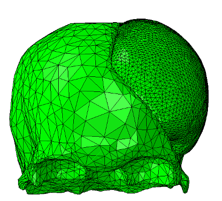
What kind of computer model is it? Not the elegant, mathematical kind of model, or the au courant 'AI' kind. Instead, I use finite element (FE) analysis, the crunchy, mechanical engineering answer to complex 3D physics problems with a range of unknowns. This is based on geometry extracted from MRI and CT scans using thresholding and existing segmentation algorithms such as FMRIB's FSL.
The extracted geometry mesh is then smoothed and optimized for FE analysis in Abaqus using commercial 3D data optimization software, 3-Matic from Materialise, and open-source software such as MeshLab.
In this case, the brain tissue itself can be modelled as hyperelastic, meaning that the deformation a material experiences is related to the force not by a simple set of constants, but instead by a function ('strain energy density' function). Typically this is relevant for applications where a material experiences a high level of deformation or has an 'unusual' structure, e.g. polymers and biological tissues.

The workings, problems, solutions of the model will be discussed in future posts. For now:
Your brain is an incredibly delicate, soft entity, with an unknown structure-function relationship to thoughts, feelings, and the broader human consciousness. Each of us carries a bowl of Jell-O, sloshing around in our heads. Yet this bowl is forming the basis of our personality, the seat of our soul, the bounds of our existence as we know it. Next time you see a kid drop some Jell-0 on the floor, spare a thought for your brain. Imagine how it responds to physical trauma, and be inspired to be careful on your commute, however you make it. Safe travels!
Back to Home Back to Technical